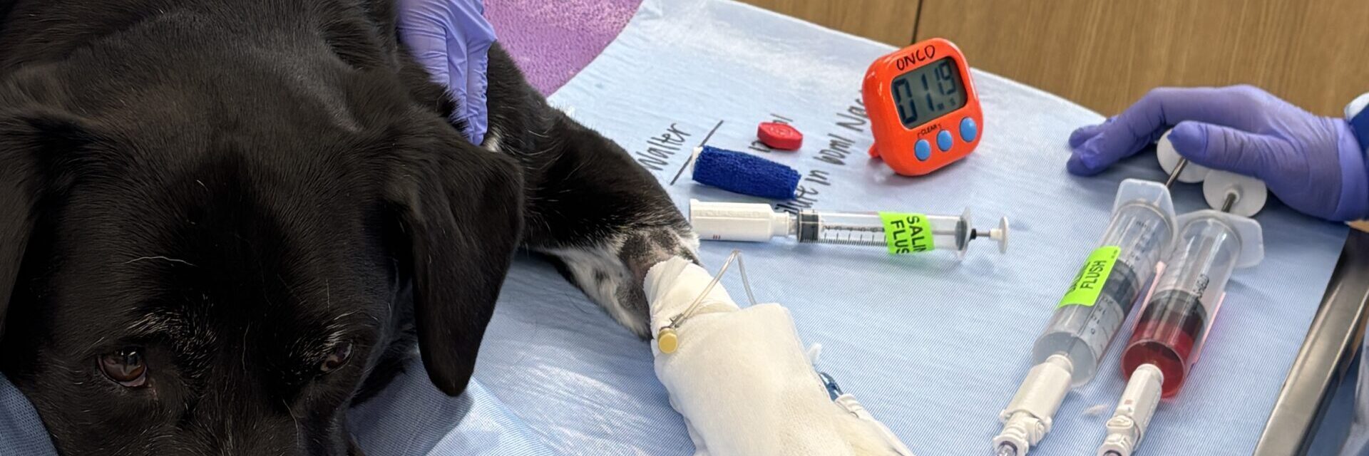
Mast cell tumors in dogs and cats: the lump that can look like anything.
Mast cell tumors are the most common skin cancer in dogs. They can look like a benign bump, a wart, a swollen area, or almost any other kind of lump — and their behavior ranges from easily cured to aggressive.
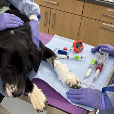
Mast cell tumors (MCTs) arise from mast cells — immune cells that normally live in the skin and mucosal surfaces and release histamine and other chemicals in response to allergens or injury. When a mast cell becomes cancerous and forms a tumor, it can carry those same chemicals with it, causing local inflammation, itching, ulceration, and sometimes systemic signs like GI upset. MCTs are the most common skin cancer in dogs and less commonly occur in cats.
How mast cell tumors typically appear.
Mast cell tumors can occur anywhere on the skin, and their appearance is famously variable. Some look like small, hairless red bumps. Others look like harmless lipomas, hairless nodules, or areas of thickened skin. Some fluctuate in size — getting bigger, then smaller — because of the histamine and inflammation the tumor cells release. That fluctuation is a hallmark sign but is not always present.
Certain breeds are at higher risk for mast cell tumors, including Boxers, Boston Terriers, Bulldogs, Pugs, Labradors, Golden Retrievers, and Shar-Peis. But any breed can develop them. In cats, mast cell tumors most often appear on the head and neck and are frequently benign.
- New skin lumps that appear over days to weeks
- Bumps that change size — getting bigger or smaller
- Red, irritated, or ulcerated areas that seem out of proportion to size
- Hairless nodules or thickened patches of skin
- In some cases, GI upset (vomiting, decreased appetite) related to histamine release from the tumor
Diagnosis and grading.
Diagnosis usually starts with a fine-needle aspirate — a quick, low-cost procedure that identifies mast cells under the microscope. Confirmed mast cell tumors are then staged to look for spread: a lymph node aspirate, abdominal ultrasound to check the spleen and liver, and bloodwork are typical parts of the workup. Some cases also benefit from CT for surgical planning.
After the tumor is removed, it is graded by a pathologist. The most common grading system uses a two-tier system (low grade or high grade). Grade matters enormously: low-grade tumors are often cured by wide surgical excision alone, while high-grade tumors often benefit from chemotherapy or other adjuvant treatment.
Treatment options.
The cornerstone of treatment is surgical removal with wide margins — typically 2–3 cm of normal tissue in every direction, plus one deep fascial plane. Getting these margins on the first surgery is one of the single most important predictors of long-term outcome.
For high-grade tumors, tumors with incomplete margins, or tumors that have spread to lymph nodes, additional treatment may include chemotherapy, targeted small-molecule inhibitors (such as toceranib, which targets the KIT mutation common in mast cell tumors), or electrochemotherapy for localized recurrences. Every treatment plan at Dogwood is tailored to the tumor’s grade, stage, and the family’s goals.
Frequently asked questions
Are mast cell tumors always cancerous?
Yes — mast cell tumors are a form of cancer. However, the behavior of a mast cell tumor varies widely by grade and location. Low-grade tumors are often cured by wide surgical excision, while high-grade tumors may need additional treatment. That’s why grading matters so much.
Can mast cell tumors spread to other parts of the body?
Yes. Mast cell tumors most commonly spread first to the local lymph node, then to the spleen and liver. Staging (lymph node aspirate, abdominal ultrasound, and sometimes bloodwork) tells us whether spread has occurred and helps guide treatment.
What does “wide margins” mean for mast cell tumor surgery?
Wide margins means removing 2–3 cm of healthy tissue around the visible tumor plus one deep tissue plane. Wide margins on the first surgery are the biggest predictor of long-term local control.
Are cats treated differently than dogs for mast cell tumors?
Yes. Feline mast cell tumors most commonly occur on the head and neck and are usually cured with surgical removal alone. Cats can also develop visceral (splenic) mast cell disease, which is treated differently.
Do I need a referral for oncology?
A referral from your primary veterinarian is helpful but not required. You can request an oncology consult directly with our team.